
Advanced Imaging
Advanced Ultrasound
Performed by specialist, Dr. Suzanne Brenner
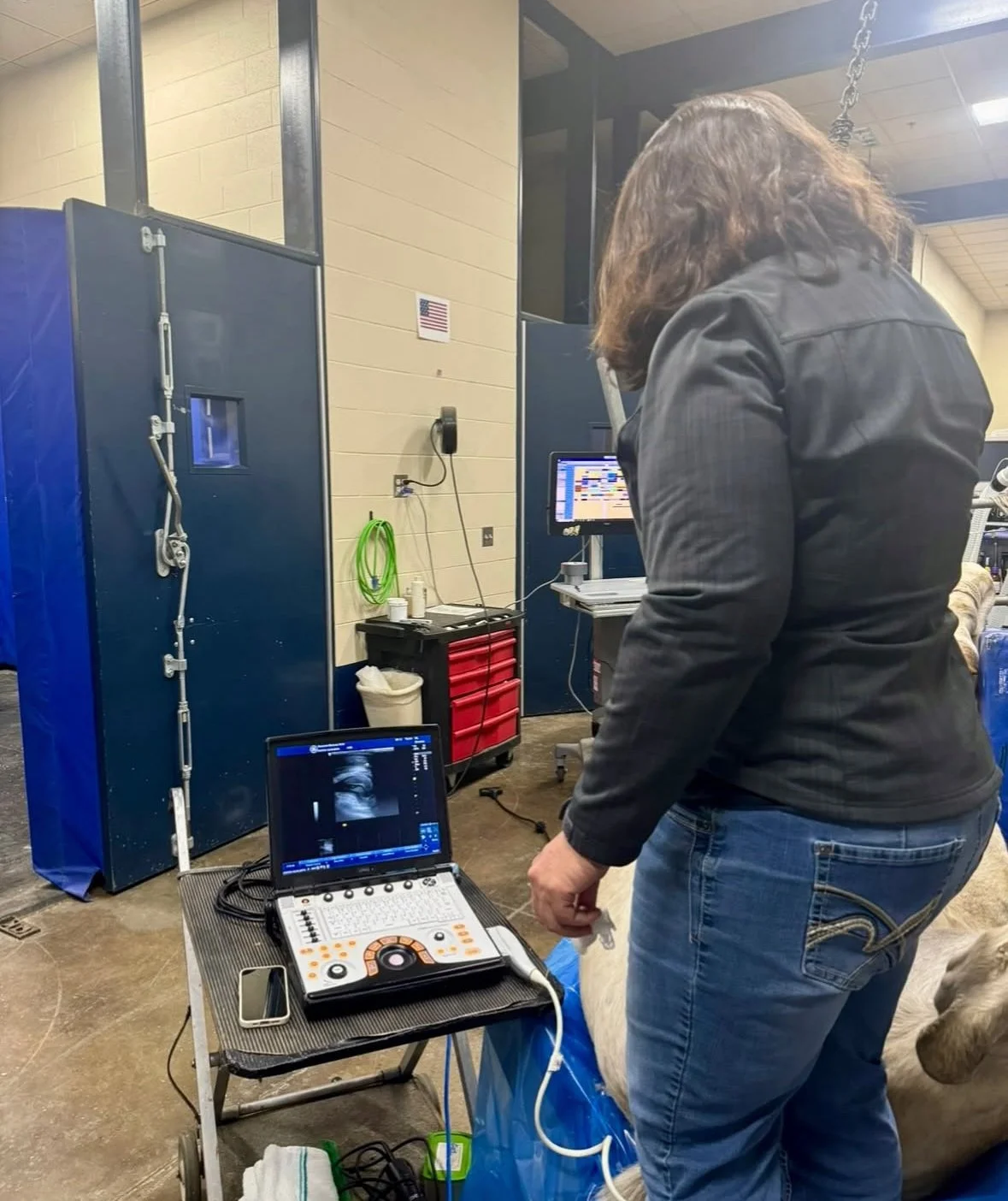
With over 20 years of experience, Dr. Suzanne Brenner is a wealth of knowledge in diagnostic ultrasound. Unlike the average practicioner, Dr. Brenner has had extensive additional training to give you maximum information whether she is scanning a limb, an abdomen, the spine, or any other part of the antanomy. Her unparalleled experience and attention to detail will get you maximal information using this routine and minimally invasive modality.
Following your lameness exam, Dr. Brenner takes her time to thoroughly ultrasound the area of concern and explain to you the findings and their implications. Dr. Brenner will work with your primary doctor, whether at Pioneer or on referral, to determine the best treatment and rehabilitation program for your horse based on their needs, injury, environement, and discipline.
Magnetic Resonance Imaging (MRI)

Our high field system allows us to acquire images in the anesthetized patient from the hoof, knee and hock; including suspicious soft tissue injuries in the pastern, fetlock, cannon, and proximal suspensory ligament areas. We gather images in multiple planes allowing the radiologist to precisely locate the cause of the problem. Though it requires general anesthesia, it provides vastly better images and more information than standing MRI units.
The superior contrast resolution of our 1.5 Telsa MRI allows the production of high quality images in any plane through the region of interest. The images generated show the anatomy of bones, joints, muscles, ligaments and tendons at a level unsurpassed by any other modality. In addition to showing normal anatomy, MRI can detect tissue injury that on gross examination may not even be apparent to the human eye. It is the most powerful tool currently available to assess the cause of lameness in horses and has revolutionized the way in which veterinary orthopedic specialists manage equine lameness.
Computed Tomography (CT)
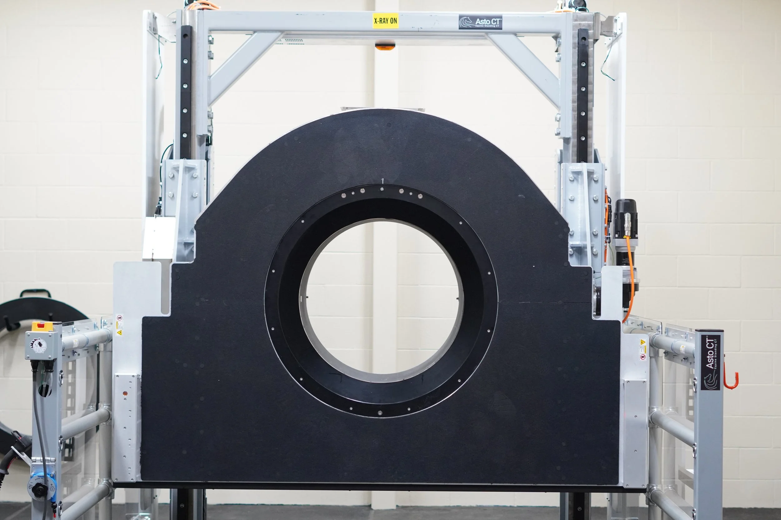
At Pioneer, we use one of the most advanced computed tomography (CT) scanners, the Asto Equina Standing CT. With this unit we are able to do scans of the head, neck, and limbs up to the knee and hock STANDING!
Computed tomography takes many x-ray measurements from different angles to produce virtual “slices” of the specific area being scanned, allowing us to see very subtle fractures, ligament tears, cartilage lesions, masses, abnormalities in the sinus and roots of the teeth.